Molecubes Modular Benchtop Preclinical PET Imaging System
Sub-millimeter image resolution is achieved through the combination of monolithic scintillators, the latest photon counting technology and GPU-based event positioning, and iterative image reconstruction.
The 5-ring configuration ensures best-in-class sensitivity over a field of view that is adequate for whole-body mouse and rat imaging at a high count rate.
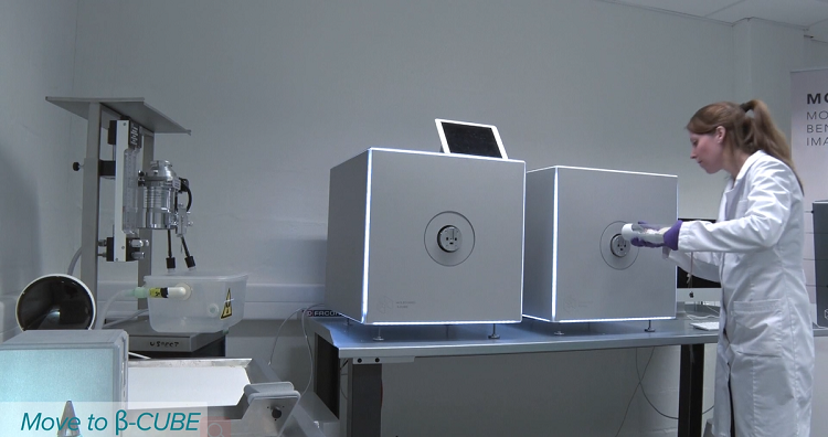
In-house hardware enables dynamic and gated studies. Intuitive, wireless acquisition software, combined with our multimodal small-animal bed, enables easy and modular multimodal imaging when used along with the γ-CUBE (SPECT) and X-CUBE (CT).
Detail
Product Principles
Positron Emission Tomography (PET)
PET is a tomography imaging device specially used for the experimental study of small animals in vivo. Compared with the clinical PET system, the spatial resolution and sensitivity of the system are higher than that of the clinical PET system, so as to adapt to the requirements of the study of small animal models. PET principle is: The developer (such as fluorodeoxyglucose, FDG) labeled with short decay period radionuclides (such as 18F, 11C, 15O, etc.), injected into the organism, releases positrons during the decay process, and a positron annihilates after encountering an electron after traveling a few tenths of a millimeter to a few millimeters. This produces a pair of high-energy gamma photons of 511KeV energy in opposite directions (180 degrees). This pair of photons is captured by a highly sensitive camera and then scattered by a computer and corrected for random information. By doing the same analysis on different positrons, we can get a three-dimensional image of how the developer accumulates in the organism. PET is the only new imaging technology that can display biomolecular metabolism, receptor and neuromediator activities in vivo, and has been widely used in the diagnosis and differential diagnosis of a variety of diseases, disease judgment, efficacy evaluation, organ function research and new drug development. It has the characteristics of high sensitivity, high specificity, whole body imaging and good safety.

Brief Introduction
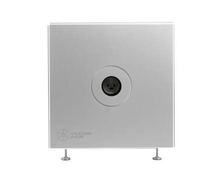
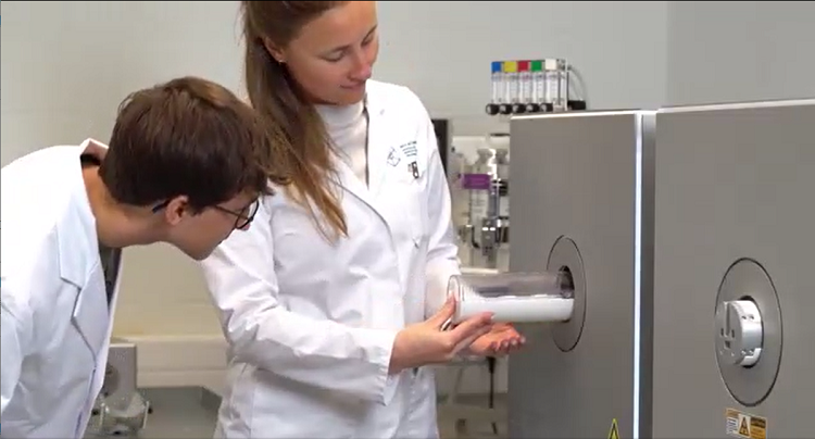
The β-CUBE is our high-performance preclinical PET imager.
Sub-millimeter image resolution is achieved through the combination of monolithic scintillators, the latest photon counting technology and GPU-based event positioning, and iterative image reconstruction. The 5-ring configuration ensures best-in-class sensitivity over a field of view that is adequate for whole-body mouse and rat imaging at a high count rate. In-house hardware enables dynamic and gated studies. Intuitive, wireless acquisition software, combined with our multimodal small-animal bed, enables easy and modular multimodal imaging when used along with the γ-CUBE (SPECT) and X-CUBE (CT).


Product Characteristics
Highest Throughput – Fast and sensitive/low dose
High-end Performance – PET/CT and SPECT/CT without compromise
Compact – light and mobile
Ease of use – functional and straight-forward
Best Uptime in preclinical industry
Fast workflow
Thanks to our fast and simple CUBEFLOW and intuitive user interfaces, 3D images are obtained using a turnkeysolution.
High precision
Extremely high-quality imaging in terms of resolution and precision and field of view at the smallest footprint.
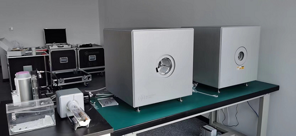
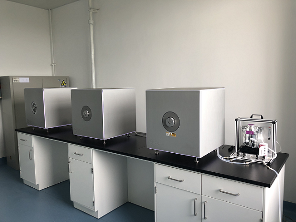
Modular
Our CUBES are designed to work bothas stand-alone units and in modular combinations so you can optimize yourworkflow and increase throughput.
Benchtop
MOLECUBES offers exquisite whole body mouse and rat imaging while fitting on top of any classic lab bench.
Best uptime
98,6% System uptime and a dedicated support team with extensive preclinical imaging expertise.
User friendly
Developed by end users, we pride ourselves in our intuitive software that allows even the most novice users tocapture beautiful images.

Application Fields
1. Oncology research: tumor genesis, metabolism, apoptosis, lymphatic vessel imaging, hypoxia imaging, tumor receptor research, tumor gene imaging, tumor treatment evaluation, etc.;
2. Research on neurological diseases: brain perfusion imaging, brain receptor imaging, brain metabolism imaging, neurodegenerative diseases (Alzheimer's disease, Parkinson's disease), inflammatory molecular imaging, etc.;
3, cardiovascular system disease research: heart disease (coronary heart disease, heart failure, heart transplant evaluation), atherosclerosis, thrombosis, cardiac nerve receptor research, etc.;
4. Gene therapy imaging: molecular imaging evaluation of gene therapy;
5, cell tracer technology: stem cell therapy, all kinds of diseases (nervous system diseases, myocardial infarction, etc.) after cell therapy evaluation;
6, new drug research and development: preclinical experiments (biological distribution, pharmacokinetics), drug target confirmation, drug lead compound screening;
7. Others: bone disease research, infectious disease research, metabolic disease research, etc.
Application
1、High resolution PET imaging
PET has high resolution: (1) The zebrafish is imaged in a semi-solid hydrogel, keeping it alive and restricting movement. The zebrafish is only 1-2cm long and its spine can be seen clearly. (2) The tumor on the alveoli of mice (about 0.5mm) can be clearly seen; (3) The striatum of the mouse brain (about 2-3mm) can be clearly seen; (4) The fine structure of the mouse heart, such as the mastoid muscle (about 1mm), can also be seen clearly. PET has a high resolution.

2、Very low dose PET imaging
Because of its high resolution and high sensitivity, PET can be imaged at extremely low radiation doses (which only means high performance, and are not possible with such low doses in practice).

3、Whole body PET imaging of one-bed mice
PET single bed FOV can be used for whole-body imaging of whole mice, and high-throughput imaging can be used for simultaneous imaging of 4 mice.

4、PET imaging for Oncology
In the [18F]FDG PET-PDAC mouse model, the tumor lesion was characterized by high uptake of [18F]FDG. The study of pancreatic tumors also revealed kidney metastases. 12.11 MBq (327 µCi) FDG, 10min PET.

5、Imaging CAR T Cell Trafficking
The researchers developed positron emission tomography (PET) probes based on the small-molecule antibiotic trimethoprim (TMP) that can be used to image the expression of the Escherichia coli dihydrofolate reductase enzyme (eDHFR) and tested the ability of [ 18F]-TMP, a fluorine-18 probe, to image primary human chimeric antigen receptor (CAR) T cells expressing the PET reporter gene eDHFR, yellow fluorescent protein (YFP), and Renilla luciferase (rLuc).

6、Dynamic PET imaging of drug metabolism
Drug metabolism study: 20min dynamic PET/CT images showed that the drug was toxic to the liver and could not remain in the liver. The liver was not bright at the end of the dynamic imaging, indicating that there was no drug residue. Dynamic 20 minute PET acquisition, 4.44 MBq (120 µCi) @ start acquisition.

7、ECG-gating PET imaging
PET images of cardiac systole (ES) and diastole (ED) in rats. The structure of the heart is clearly seen in the two images on the right. Mice are similar.

8、Bone PET imaging
highly sensitive bone-friendly PET imaging agent used to detect bone lesions and to view bone diseases. The bright tips of the bones are growth plates. 1.74 MBq (47 µCi) @start acquisition, 10 min PET acquisition.

Video
Molecubes Introduction 1

Molecubes Introduction 2