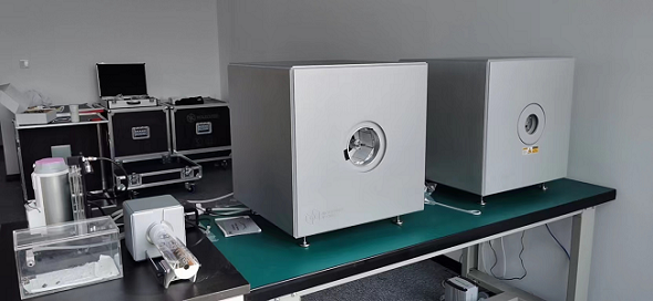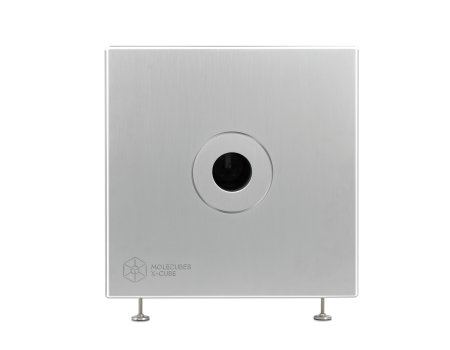
Molecubes Modular Benchtop Preclinical CT Imaging System
The light weight of this self-shielded imaging unit makes it a truly mobile in-vivo scanner.
Advanced workflows, such as gated and dynamic contrast enhanced imaging, can be achieved in a functional and integrated setup.
Our iterative reconstruction techniques are available in standard as well as in expert user mode.
Intuitive, wireless acquisition software, combined with our multimodal small-animal bed, enables easy and modular multimodal imaging when used along with the γ-CUBE (SPECT) and β-CUBE (PET).
Detail
Product Principles
Computed Tomography (CT)
Small animal CT uses X-ray beam to scan the layer of a certain thickness in a certain part of the organism. According to the different absorption and transmittance of X-ray in different tissues, the X-ray through the layer is received by the detector, converted into visible light, converted from photoelectric to electrical signal, and then converted into digital by analog/digital converter, input computer processing. After the data is processed by the computer, the cross-section or three-dimensional image of the part of the organism being examined can be taken, which is especially suitable for imaging parts such as bones.

Brief Introduction
The X-CUBE is our high throughput CT “work horse”. It allows for fast whole body mouse and rat CT imaging at extremely low dose and excellent soft tissue contrast.
The light weight of this self-shielded imaging unit makes it a truly mobile in-vivo scanner. Advanced workflows, such as gated and dynamic contrast enhanced imaging, can be achieved in a functional and integrated setup. Our iterative reconstruction techniques are available in standard as well as in expert user mode. Intuitive, wireless acquisition software, combined with our multimodal small-animal bed, enables easy and modular multimodal imaging when used along with the γ-CUBE (SPECT) and β-CUBE (PET).


Product Characteristics
Highest Throughput – Fast and sensitive/low dose
High-end Performance – PET/CT and SPECT/CT without compromise
Compact – light and mobile
Ease of use – functional and straight-forward
Best Uptime in preclinical industry
Fast workflow
Thanks to our fast and simple CUBEFLOW and intuitive user interfaces, 3D images are obtained using a turnkeysolution.
High precision
Extremely high-quality imaging in terms of resolution and precision and field of view at the smallest footprint.
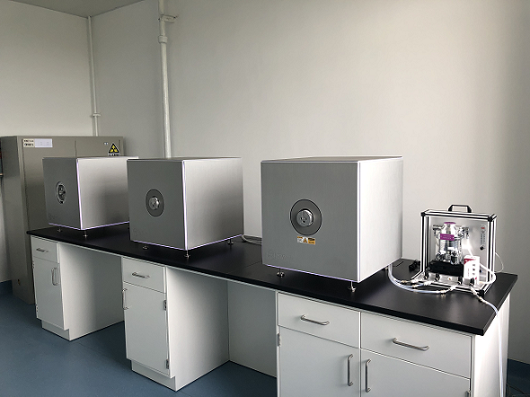
Modular
Our CUBES are designed to work bothas stand-alone units and in modular combinations so you can optimize yourworkflow and increase throughput.
Benchtop
MOLECUBES offers exquisite whole body mouse and rat imaging while fitting on top of any classic lab bench.
Best uptime
98,6% System uptime and a dedicated support team with extensive preclinical imaging expertise.
User friendly
Developed by end users, we pride ourselves in our intuitive software that allows even the most novice users tocapture beautiful images.
Very low dose imaging (noise reduction techniques and iterative reconstruction algorithms) :

Contrast-enhanced CT:

Application Fields
Application objects of Molecubes CT:
(1) in vivo: The research objects are living mice and rats, which can realize the longitudinal study of physiological and metabolic functions. Similar to clinical CT, small animal CT can also perform respiration/heartbeat gated and developer enhanced scanning.
(2) in vitro: The research object is in vitro specimens (such as bone, teeth) or samples of various materials. A coagulated contrast agent can also be used to infuse live animals for fine imaging of the cardiovascular, urinary, or digestive systems.
Main applications of Molecubes CT:
1. Bone: Bone research is one of the most important applications of preclinical CT because of the relatively obvious difference between bone and other body tissues in X-ray attenuation performance. Cortical bone and bone trabecular analysis can be performed, and the changes are associated with conditions such as osteoporosis, fractures, osteoarthritis, ischemia, and genetic diseases, as well as bone tumors.
2. Teeth and periodontal tissues: From the 3D overall structure, root canal morphological changes, dental caries destruction, dental tissue density changes, alveolar bone structure and mechanical properties changes can be studied.
3. Biomaterials: Analyze the porosity, strength and other parameters of biomimetic materials prepared in vitro; Scan tissue samples that need to be replaced to obtain three-dimensional images; Imaging analysis of medical device materials, such as heart stents, cerebrovascular stents, etc. Implant compatibility analysis; Let's wait.
4. Disease Detection and Research:
(1) Oncology research: It can study the occurrence and treatment evaluation of lung tumors, brain tumors and parenteral tumors; CT can detect the nature of the tumor, such as the growth form of the tumor, the degree of invasive growth, and whether the surrounding boundary is clear and whether there is a complete envelope.
(2) Bone research: Study the influence of different genes or signaling pathways on the quantity or quality of bone, the influence of disease states on bone development/repair, the influence of cytokines on blood vessel growth during tissue repair after fracture, etc.
(3) Brain research: It is mainly used to diagnose cerebral vascular lesions and intracranial hemorrhage, and the examination does not necessarily need to use a developer. In the case of acute stroke, the possibility of bleeding can be ruled out. In addition, it is also very useful in the diagnosis of skull and facial bone fractures with trauma.
(4) Lung research: In the diagnosis of lung tissue, there is a high diagnostic value for acute or chronic changes, such as pneumonia or tumors. Some interstitial tissue changes (lung parenchyma, lung fibers, etc.) can be reconstructed with high resolution Settings of thin sections.
(5) Angiography: The developer is rapidly injected into the heart cavity or blood vessel through the cardiac catheter, so that the heart and blood vessel cavity can be developed under CT irradiation. Using contrast media CT, the vascular system and blood-rich tissues and organs can be shown. Ranging from arteries to heart, lungs, kidneys, liver, etc. Provides a better soft tissue contrast for sharper imaging.
(6) Heart research: The data of cardiac tomography can be combined with gated observation of the heart beat to reduce the moving false image caused by high-speed heartbeat, and can also preliminarily check the health status of the heart muscle.
(7) Analysis of body fat distribution: study the distribution rule of adipose tissue, redistribution under certain pathological conditions (such as Cushing syndrome, etc.), the relationship between obesity and metabolism-endocrine disorders and cardiovascular system complications.
(8) Inflammation, calcification (to evaluate the effect of hyperlipidemia on heart valve calcification), stones.
(9) Nanotracers: Research on applications related to novel nanotracers in the fields of urology, thyroid, and oncology, especially the transport mechanisms of nanoparticles, such as the different transport processes and distribution characteristics of gold nanoparticles metabolized by the kidney in normal and injured kidneys.
5. New drug development: research on new osteoporosis drugs and efficacy evaluation, etc.
6, auxiliary positioning: combined with preclinical PET, SPECT, or with small animal live optical imaging, can be used for structural imaging, auxiliary positioning.
7. Others: Structural detection of other organic or inorganic materials, quality inspection and flaw detection of micro-devices, fossil structure analysis, etc.
Application
1、CT imaging combined with PET
PET/CT combination images: Drug metabolism study: 20min dynamic PET/CT images showed that the drug was toxic to the liver and could not remain in the liver. The liver was not bright at the end of the dynamic imaging, indicating that there was no drug residue. Dynamic 20 minute PET acquisition, 4.44 MBq (120 µCi) @ start acquisition.

2、CT imaging combined with SPECT
SPECT/CT combination images, bone imaging of mice: 99mTc-HDP, 94.35MBq (2.55mCi) @start acquisition, acquisition time of 60min.

3、CT imaging combined with optical imaging
CT images can be fused with live optical images of small animals for structural imaging and localization.

4、Bone CT imaging
Bone tumor studies: In vivo CT in rats showed that the tumor caused brain and bone metastases, a large number of cavities in the bone, FDK reconstruction, 50µm voxel size.

In vivo measurement of rat tail vertebrae: high resolution ring acquisition, acquisition time: 2min3s, ISRA reconstruction, 50µm voxel size.

In vivo measurement of spinal vertebrae in rats: high definition ring acquisition, acquisition time: 4min, FDK reconstruction, 50 and 20µm voxel size.

Bone data processing with PMOD software: By selecting the region of interest (ROI) on the CT scan image for threshold segmentation and other operations, cortical bone (bone dense) and bone trabeculae (bone cancellous) can be segmulated and extracted into different tissue regions respectively, so as to study and analyze various morphological characteristics of them.

5、ECG/respiratory gated CT imaging
ECG gating: live mice were injected with the development enhancer Exitron 12000, with ultra-high temporal resolution (10ms), and the imaging time was 2min40s.

Respiratory gating: Ultra-high temporal resolution (10ms), 4min, 390mGy. Left: Integrated picture. Middle image: at the end of the exhalation and right image: at the end of the inhalation show a 34 percent difference in the volume of the lungs.

6、Contrast-enhanced tumor CT imaging
Tumor study: Contra-enhanced CT was used to examine liver tumors in living mice: Exitron 12000 was injected, FDK was reconstructed, and the imaging time was 4min, 100µm voxel size.

7、CT imaging of COVID-19
Detection of lung CT images of living Syrian hamsters: Qualitative analysis method: the degree of lesion was scored according to CT images; Quantitative analysis method: By analyzing the image parts with pixel density in the range of 56-255, the non-aerated lung volume on CT images was quantitatively analyzed. By analyzing the parts of the image with pixel density in the range of 0-55, the airways and aerated lung volume were quantitatively analyzed. The images showed that the yellow arrows indicated lung consolidation and the blue arrows indicated bronchiectasis.

Video
Molecubes Introduction-1